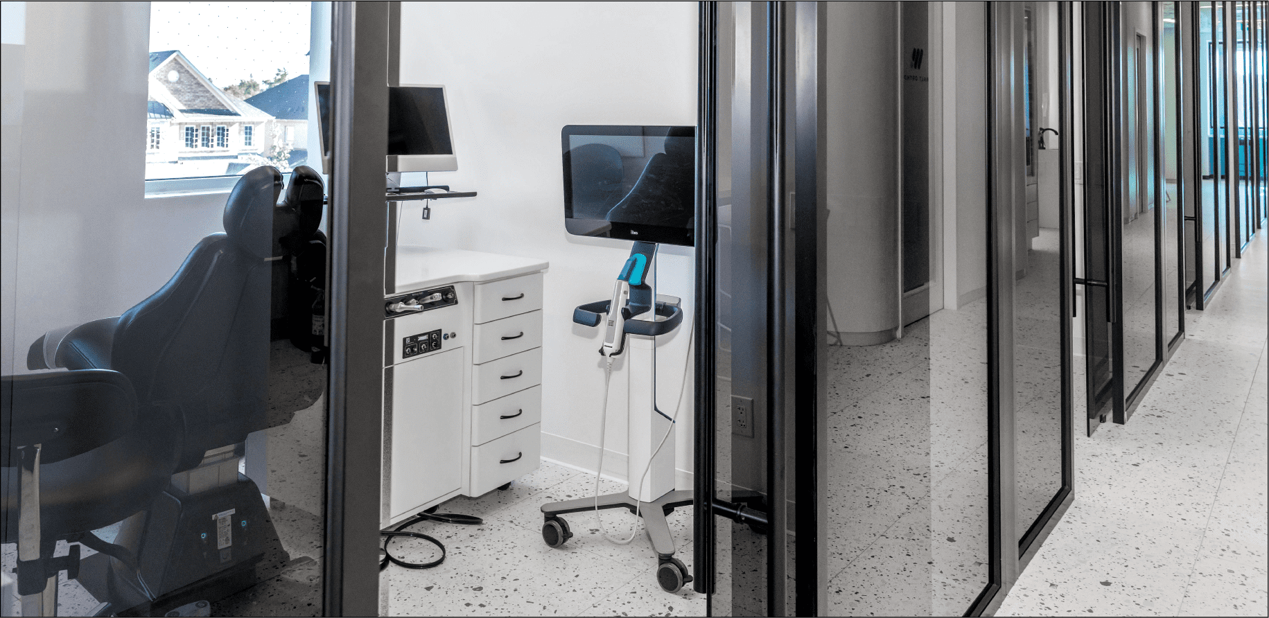
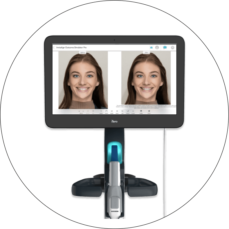
3D IMPRESSIONLESS SCANNING

AT WALT ORTHODONTICS, WE BELIEVE IN OUR PATIENTS COMFORT AND CONVENIENCE WHICH IS WHY WE ARE PROUD TO ANNOUNCE THE ADDITION OF 3D DIGITAL SCANNING IN OUR OFFICE.

The technology is so precise that often times this leads to more successful ortho cases and outcomes because the 3D rendering of your teeth is very accurate allowing for a snugger fit of the Invisalign aligners throughout your orthodontic treatment plan. Also patients love that they don’t need to take multiple impressions with the Alginate Impression material, (patients like to refer to it as goop!) To learn more about Alginate and why 3D scanning is a a better option, read our blog article “Dr Walt Making an Impression. Why 3D scanning is great.

One of the wonderful features of 3D scanning technology is that you get to see what your smile will look like at the end of the Invisalign treatment plan. This is truly a great feature of the system and is one of the reasons why we invested in the technology for our patients. It allows us to help you better visualize the results at the outset of treatment and gives you that much more reason to work with the Invisalign system.

ITERO® 3D SCANER

BENEFITS OF 3D SCANNING WITH ITERO

ACCURACY

The iTero 3D scanning system is precise resulting in better fitting aligners (if using invisalign).

COMFORT

Gone are the days of needing to subject patients to goopy impressions with alginate. iTero 3D scanning does away with messy, unpleasant impressions.

VISUALIZATION

3D scanning allows us to provide a virtual tour of what your case will look like at completion. This gives you a much better understanding of their treatment plan.

THE SCIENCE BEHIND NIRI

NIRI serves as a valuable diagnostic aid in the early detection of interproximal caries. The iTero Element Plus Imaging System uses light of wavelength (= 850nm) in the electromagnetic spectrum which, on interaction with the hard tissue of the tooth, provides additional data of its structure.

Enamel is transparent to NIRI due to the reduced scattering co-efficient of light, allowing it to pass through its entire thickness and present as a dark area, whereas the dentin appears bright due to the scattering effect of light caused by the orientation of the dentinal tubules. Any interferences/pathological lesions/areas of demineralization appear as bright areas in a NIRI image due to the increased scattering within the region.

THE BENEFIT BEHIND NIRI

Optical methods have the advantage that they do not use ionizing radiation. For this reason, these procedures can be used as often as desired to monitor caries. Several clinical studies have shown NIRI sensitivity to be as potent as radiographic examinations and are well suited for the detection and imaging of interproximal caries.

Ready for some

If you have been wanting your bespoke, dream smile, our team at Walt Orthodontics is happy to help you achieve that. Book your complimentary consultation!